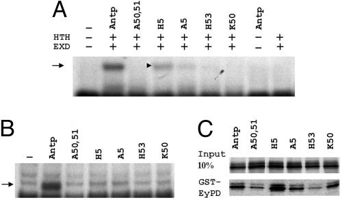
Fig. 2.
Molecular characterization of the different Antp mutant proteins. (A and B) Gel shift experiments performed on a Hox/Exd/Hth site (A) or on HB1 probe (B). Arrows indicate the specific retarded complexes. (A) Only the ternary complex is shown. ANTP does not bind on its own on this site and weakly in the presence of EXD (data not shown). (C) Coretention assay between the GST-EY-PD and the mutated ANTP proteins indicated. Input lane indicates the amount of proteins used for the assay. No binding was observed with GST alone (data not shown).

