
Fig. 1.
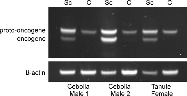
Xmrk and protooncogene expression in X. cortezi. Semiquantitative RT-PCR analyses using the same template primers for the oncogene (Xmrk, bottom bands of upper image) and protooncogene (top bands of upper image). In each of the three wild-caught individuals, all of which did not have visible melanoma formation, the Sc tissue was associated with both Xmrk and protooncogene expression, whereas in the nonpigmented control tissue (C), only the protooncogene was expressed. The housekeeping gene, β-actin, is included as a loading control (17). A 15-bp insertion associated with the protooncogene in X. cortezi accounts for the fractionation of the two bands by gel electrophoresis (A.A.F. and S. Tanda, unpublished data). This small insertion was found in the predicted signal peptide sequence at the beginning of the protooncogene. Polymorphisms within this region of the oncogene and protooncogene are common in individuals derived from wild populations of several Xiphophorus spp. (18).