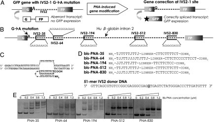
Fig. 1.
Strategy for targeted correction of beta-globin gene IVS2–1 mutation in mammalian cells. (A) Schematic of the cellular assay, in which intron 2 of the beta-globin gene, mutated at position 1, interrupts the GFP sequence. In cell lines made from this construct, this mutation leads to an aberrant transcript. GFP expression in these cells requires reversion of this IVS2–1 G→A mutation, via PNA-stimulated gene correction, thereby restoring proper splicing. (B) Relative positions and sequences of the homopurine bis-PNA binding sites in intron 2 of the beta-globin gene. (C) Putative strand invasion complex formed by bis-PNA-35 at the beta-globin IVS2–35 site, resulting in a PNA/DNA/PNA triplex structure. (D) Sequences of the bis-PNAs used in the current study. The sequence of the 51-mer IVS2 donor DNA corresponds to the nontemplate strand (single base pair change underlined). J, pseudoisocytidine. (E) Gel shift assays of the bis-PNAs binding to their respective target sites in a plasmid construct.

