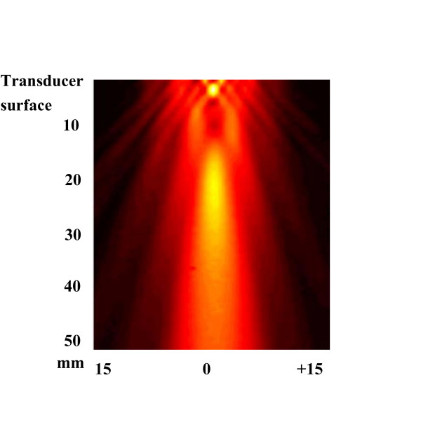
Figure 1.
The field distribution for the transducer used in the present study. Needle hydrophone exploration of field distribution for the transducer. Scanning was performed over an area of 50 × 30 mm2 in the y- and z-direction starting close to the transducer surface. No exact values of intensity were measured. Clots were placed 30 mm from the transducer surface.

