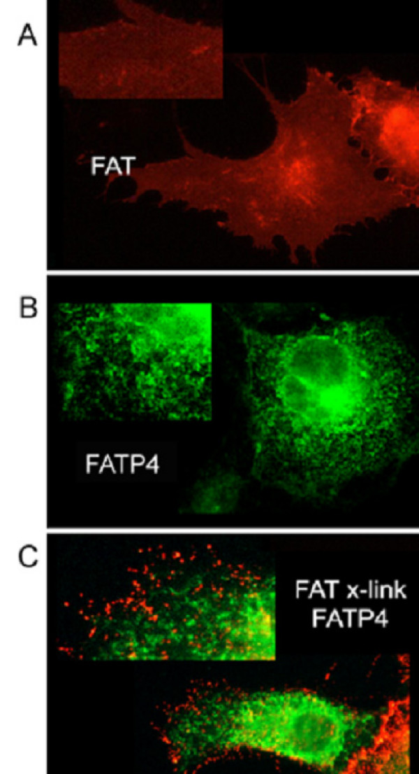
Figure 4.
FAT/CD36 does not co-localize with FATP4. 20 h after transient transfection with FAT/CD36 and FATP4-GFP, FAT/CD36 was clustered with anti-FAT/CD36 antibody from Biosource and Cy3 fluorescently labelled secondary antibody. (A) FAT/CD36 is mainly localized at the plasma membrane. (B) FATP4-GFP shows a reticular staining pattern as described before [13] representing ER membranes. (C) Patched FAT/CD36 does not show any co-localisation with FATP4-GFP.

