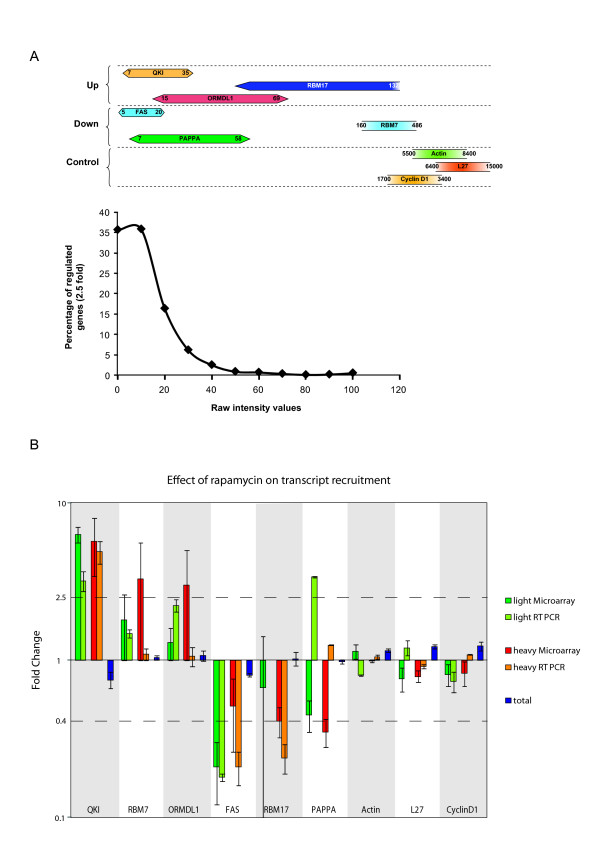
Figure 3.
Real-Time RT-PCR analysis. (A). A schematic representation of the intensity values of transcripts selected for the RT-PCR validation. In the upper panel, the horizontal bars represented the value range for each mRNA on the array. The lower panel plots the distribution of regulated genes (×2.5 fold cut-off selection) relative to the probe set values. (B). RT-PCR values were normalised to those obtained from two housekeeping genes and the fold change indicates the difference in the DMSO and rapamycin values after normalisation. The DMSO value was arbitrarily set at 1. The results are compared with those obtained from the microarray. The variation in the total mRNA extracted is also represented. The 2.5 fold difference used for the screening is represented by the dotted lines (2.5 and 0.4). Each bar is representative of 2 independent RT-PCR assays performed in triplicate. Bars indicate the SEM.

