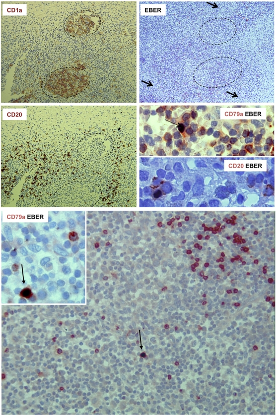
Figure 2. Detection of EBV-infected B cells by in situ hybridization combined with immunohistochemistry.
Granuloma serial sections were stained for CD1a (upper left) or for CD20 (middle left) by immunohistochemistry or for EBERs by in situ hybridisation (upper right, arrows indicate EBER positive cells). Combined detection of CD20 or CD79a with EBERs shows that EBV-infected cells are B cells (arrows).

