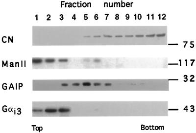
Figure 2.
Distribution of GAIP in AtT-20 cell fractions prepared by sucrose density centrifugation as described in MATERIALS AND METHODS. The ER marker calnexin peaks in heavy fractions with the majority (71%) in fractions 9–12. The Golgi marker Man II and Gαi3 are found in light fractions 1–3, which also contain PM. Fractions 1–3 contain 15, 44, and 40%, respectively, of the total Gαi3 in the gradient. GAIP is associated with fractions 3–7 of intermediate density, which have 14, 17, 38, 17, and 13%, respectively, of the total. Thus, the overlap between the distribution of GAIP and that of Gαi3 is limited to fraction 3 and is minimal (14%). Fractions were lysed in Laemmli buffer, and proteins from each fraction were separated by SDS-PAGE, transferred onto PVDF membranes, reacted with anti-GAIP (C) antiserum (diluted 1:2000) followed by goat anti-rabbit IgG coupled to HRP (1:3000), and immunoreactivity was detected by chemiluminescence (ECL).

