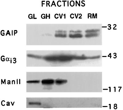
Figure 7.
Distribution of GAIP in fractions prepared from rat liver. GAIP is concentrated in fractions enriched in carrier vesicles (CV1 and CV2). It is not detected in Golgi light (GL) fractions and is barely detectable in Golgi heavy (GH) fractions. Rat liver was homogenized and fractions were prepared by sucrose gradient centrigugation as described in MATERIALS AND METHODS. Fifty micrograms each of Golgi light (GL), Golgi heavy (GH), carrier vesicles 1 and 2 (CV1 and CV2), and residual microsome (RM) fractions were solubilized in Laemmli buffer and immunoblotted for GAIP (N-terminal antibody, dilution 1:4000), Gαi3 (EC antibody, dilution 1:1000), Man II (dilution 1:2000), and caveolin (dilution 1:4000) as in Figure 1. The presence of two bands for GAIP is likely due to posttranslational modification (palmitoylation or phosphorylation).

