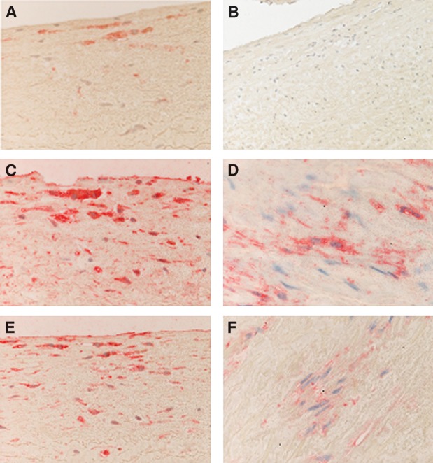
Fig. 1.
Immunohistochemical analysis of atherogenic aortas. A: CD68 expression in macrophages located in the intima; original magnification × 200. B: Staining with anti-CD3 showing the absence of T cells and serving as a blank; original magnification × 200. C: PAF-acetylhydrolase (PAF-AH) cytoplasmic expression in macrophages; original magnification × 200. D: PAF-AH expression in smooth muscle cells (SMCs); original magnification × 400. E: PAF receptor (PAF-R) expression in monocyte-macrophages in the intima; original magnification × 200. F: PAF-R expression in SMCs; original magnification × 400.

