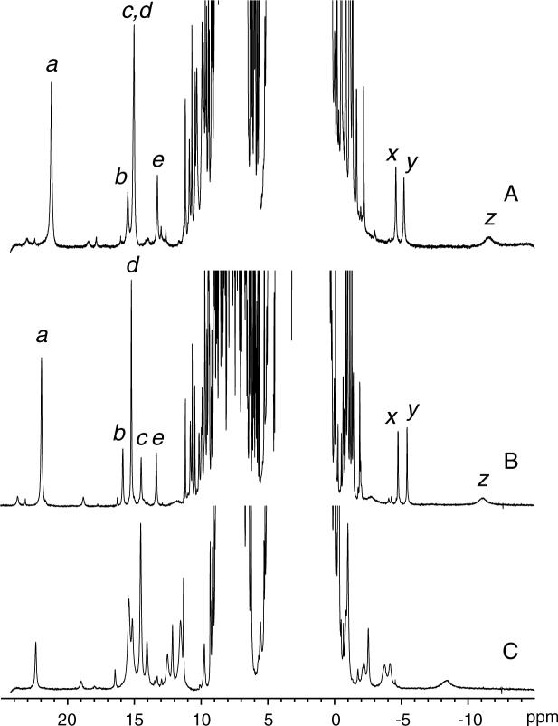
Figure 3.
1H spectrum of (A) ferric wild-type S6803 rHb-R, (B) ferric A69S S6803 rHb-R, and (C) cyanomet A69S S6803 rHb-R. Data were collected at 25 °C, pH 7.2. In traces A and B, the labels are as previously used (4, 41): a, heme 5-CH3; b, heme 2-α vinyl; c, His70 NδH; d, heme 1-CH3; e, His46 NδH; x, y, 2-β vinyls; z, His46 CεH. The resonance at 22.5 ppm in trace C arises from Tyr22 OηH hydrogen-bonded to the cyanide ligand.

