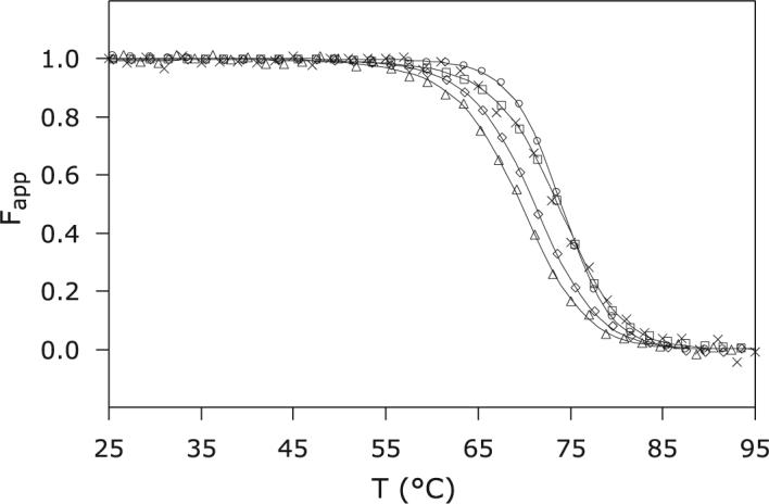
Figure 4.

Thermal denaturation of S6803 rHb-R. CD and visible spectroscopy data were collected at pH 7.2 on the wild-type and A69S proteins in the ferric state. ×: Ferric wild-type S6803 rHb-R, CD; □: ferric wild-type S6803 rHb-R, visible; Δ ferric A69S S6803 rHb-R, CD; △: ferric A69S S6803 rHb-R, visible; ○: cyanomet A69S rHb-R, visible.
