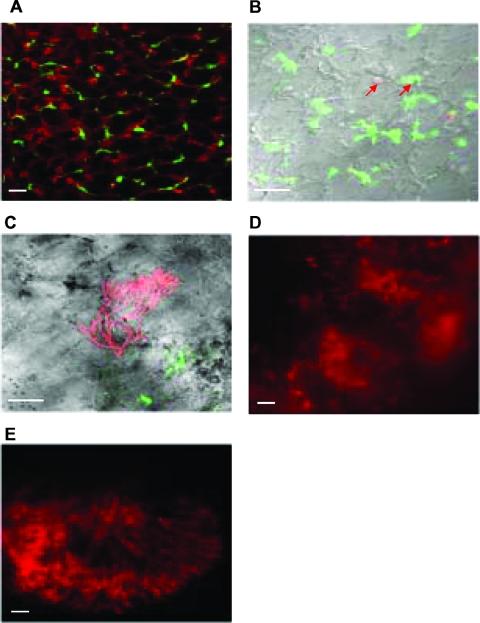
FIG. 1.
Ex vivo confocal imaging of cross sections of fresh lung tissue from CX3CR1GFP mice. (A) Lungs were injected with CMTMR to reveal tissue architecture. Mice were intranasally infected with 107 DsRed A. fumigatus conidia, and the course of infection was observed for immunocompetent mice at day 4 (B) and for immunosuppressed mice at day 4 (C and D) and at day 7 (E). (B) Examples of DsRed A. fumigatus phagocytosed by MΦ and DC are indicated by the red arrows. Hyphal forms are observed starting on day 4 in immunosuppressed mice (C, D, and E). Scale bars = 10 μm.

