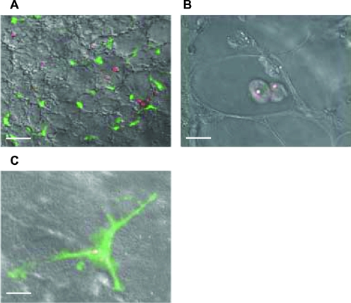
FIG. 3.
Confocal microscopic analysis of the lungs of C16-KAAK-treated immunocompromised CX3CR1GFP mice. Four days after mice were intranasally infected with DsRed A. fumigatus conidia, fresh lung sections were isolated. Residual conidia were detected within both MΦ (A and B) and DC (A and C). Scale bars = 10 μm (A) and 5 μm (B and C).

