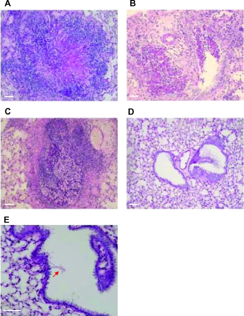
FIG. 5.
Fungus detection on lung sections of infected immunocompromised WT mice. Lungs of mice from each group of the second survival assay were isolated at the last survival day. Lung paraffin sections were stained with PAS. Focal lesions of IA were observed in both untreated (A and B) and AMB-treated (C) mice. Lung tissue of C16-KAAK-treated mice, at 21 days after infection, was cleared of fungal forms (D), and only a few damaged hyphae were found as indicated by the red arrow (E). Scale bars = 10 μm.

