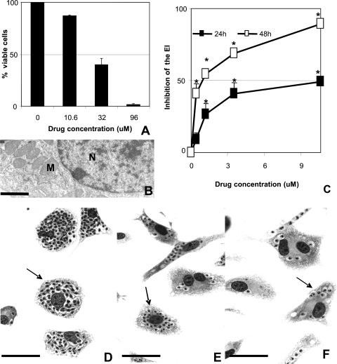
FIG. 3.
Cytotoxicity analysis of DB1362 in mammalian host cells (A and B) and diamidine activity against T. cruzi-infected peritoneal macrophages after 24 (C) and 48 (C to F) h of treatment. The effect of DB1362 on mammalian host cells was evaluated by both trypan blue exclusion assays (A) and TEM (B). Loss of cellular viability was noticed only when the cultures were incubated with 32 μM or higher doses (A). Ultrastructural analysis shows characteristic nuclei and mitochondria of the mammalian cell after exposure to the diamidine (B). N, nuclei; M, mitochondria. Bar = 10 μM. (C to F) DB1362 activity against intracellular amastigotes in T. cruzi-infected host cells. The activity of DB1362 after 24 and 48 h of drug incubation is shown by the inhibition of the endocytic index (EI) (C). Asterisks indicate significant differences between treated-T. cruzi-infected peritoneal macrophages and untreated cultures (P ≤ 0.05). (D to F) Light microscopy of untreated (D) and T. cruzi-infected (E and F) host cells exposed to 0.39 μM (E) and 1.18 μM (F) DB1362 for 48 h. Arrows indicate intracellular parasites. Bars = 1 μm.

