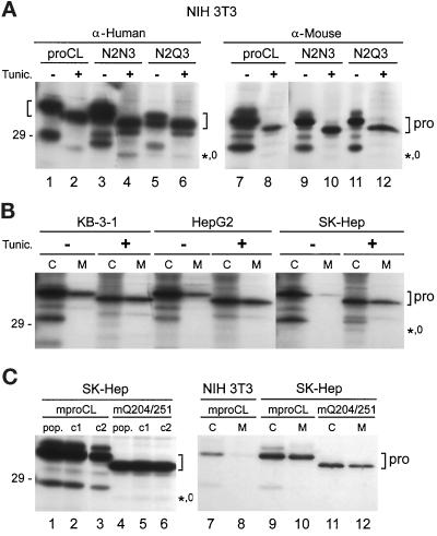
Figure 8.
Tunicamycin treatment. (A) Transfected cell lines were incubated in the presence (+) or absence (−) of tunicamycin (tunic.) for 4 h and then radiolabeled for 3 h with [35S]methionine (+/- tunic.). Cell lysates were immunoprecipitated with anti-human proCL antiserum or anti-mouse proCL antiserum, as indicated, followed by SDS-PAGE and fluorography. (B) Three human cell lines, as indicated, were treated with tunicamycin (+) or left untreated (−) and radiolabeled with [35S]methionine. Cell lysates (C) or media (M) were immunoprecipitated with anti-human proCL antiserum, followed by SDS-PAGE. (C) Human SK-Hep cells were transfected with wild-type mouse proCL (mproCL) or with a mutant lacking both of its normal glycosylation signals (mQ204/251). Left, Populations of cells (pop.) or two individual clones from each population (c1 and c2) were radiolabeled and cell lysates were immunoprecipitated with anti-mouse proCL antiserum. Right, NIH3T3 cells expressing authentic mouse proCL or populations of SK-Hep cells expressing wild-type or mutant recombinant mouse proCLs, as indicated, were radiolabeled with [35S]methionine. Cell lysates (C) and media (M) were immunoprecipitated with anti-mouse proCL antiserum. Brackets mark the positions of proenzymes (pro). The positions of the small amount of unglycosylated, lysosomal proteins are indicated (*,0). The 29-kDa size standard is shown on the left and lane numbers are provided below the panels.

