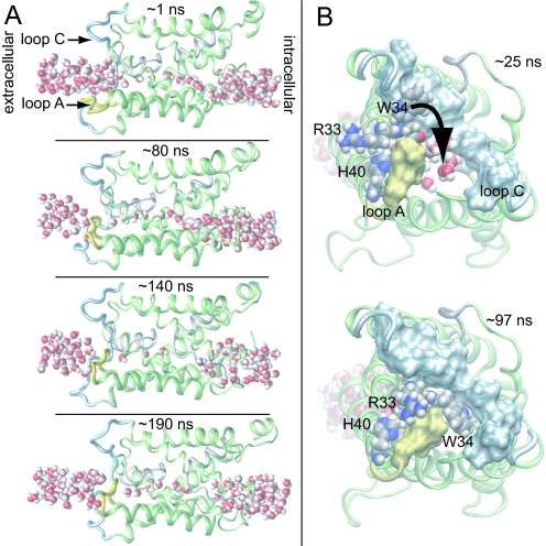
Fig. 4.
Channel occlusion. (A) Consecutive snapshots from the AQP0NJ4 simulation. The water pathway is pinched off by intrusion of loop A (yellow) while loop C (light blue) remains stationary. The lumen dries out on occlusion, but rehydrates later in the simulation (Lower Middle and Bottom) after loop A reopens. (B) Extracellular view. His-40, Arg-33, and Trp-34 are shown as spheres. Trp-34 slides between loops A and C, thereby occluding the channel.

