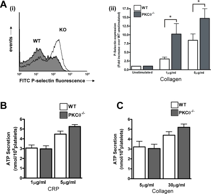
Figure 3. PKCθ negatively regulates α-granule secretion.
A: Washed platelets from WT or KO mice were stimulated with CRP (1 or 5 µg/ml) in the presence of FITC-labelled anti-P-selectin antibody for 15 minutes, and surface labelling measured by flow cytometry. Representative histograms are shown in A(i) (1 µg/ml), and fold-increase in geometric mean compared to unstimulated platelets is shown in A(ii) (mean±SEM; n = 8). B, C: ATP secretion from dense granules in response to CRP (B) or collagen (C) was monitored in a luminometer using the luciferin-luciferase reaction. Data are presented as mean±SEM (n = 4).

