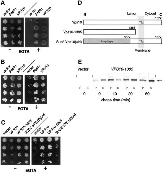
Figure 4.
Suppression of EGTA hypersensitivity in pmr1 cells by the sorting receptor Vps10 is mediated through its luminal domain. (A) Suppression by wild-type Vps10. Serial fivefold dilutions of saturated cultures were spotted onto complete medium lacking uracil. Addition (+) or omission (−) of 6.5 mM EGTA is indicated. Plates were photographed after 3 d incubation at 30°C; strains are (left to right): YR439 (pmr1, 2μ-vector), YR657 (pmr1, 2μ-PMR1), and YR480 (pmr1, 2μ-VPS10). (B) Vps10-mediated suppression does not require Pmc1. Conditions are as in panel A. Strains (from left to right): YR469 (pmr1 pmc1 cnb1, 2μ-vector), YR472 (pmr1 pmc1 cnb1, 2μ-PMR1), and YR477 (pmr1 pmc1 cnb1, 2μ-VPS10). (C) The luminal domain of Vps10 is necessary and sufficient for suppression. Same conditions as in panel A; strains are (from left to right): YR547 (pmr1, 2μ-vector), YR551 (pmr1, 2μ-VPS10), YR550 (pmr1, 2μ-VPS10–1385), and YR549 (pmr1, 2μ-SUC::VPS10-ΔN). (D) Vps10 derivatives used in the analysis. The transmembrane topology of Vps10 and its derivatives is depicted; numbers indicate the Vps10 residues present in each derivative. (E) Vps10–1385 is secreted by pmr1 cells during growth in EGTA-containing medium. Cells were cultured in synthetic complete medium, converted to spheroblasts, and labeled (15 μCi/OD cells) with 35S-methionine as described (Horazdovsky and Emr, 1993), but EGTA (3 mM) was present during these steps. At the times indicated, cells and media supernatants were analyzed for the presence of Vps10–1385 by immunoprecipitation, SDS-PAGE, and autoradiography. The band corresponding to Vps10–1385 in SDS-PAGE is marked (arrow). Strains: YR547 (pmr1, 2μ-vector) and YR550 (pmr1, 2μ-VPS10–1385).

