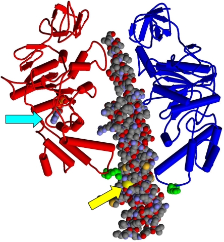
FIGURE 1.
The 380βDELSEED386 motifs relative to the catalytic sites. Two of the β subunit conformers, βDP (red) and βE (blue), and the coiled-coil region of the γ subunit (space-filling representation) from the x-ray structure of Abrahams et al. (69) are shown. ADP bound to βDP is shown in the space-filling model (cyan arrow). βAsp-380 and βGlu-381 (E. coli numbering) are shown in green space-filling representation near the “bottom” of the β subunits in the βDELSEED loop. γCys-87 is indicated by the yellow arrow.

