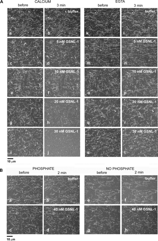
FIGURE 2.
GSNL-1 severs actin filaments. A, Alexa488-labeled actin filaments were tethered to a glass coverslip and treated with buffer alone (a, b, k, and l) or varying concentrations of GSNL-1 in the same buffer (c–j and m–t) for 3 min. The buffer in a–j contained 0.1 mm CaCl2, and the buffer in k–t contained 5 mm EGTA. The filaments were observed before (a, c, e, g, i, k, m, o, q, and s) and 3 min after the incubation (b, d, f, h, j, l, n, p, r, and t), and micrographs of the same fields were taken. Bar, 10 μm. B, Alexa488-labeled actin filaments were preincubated with buffer containing 10 mm potassium phosphate (a and c) or no phosphate (e and g). Then buffer alone containing 10 μm CaCl2 and no phosphate (b and f) or the same buffer containing 40 nm GSNL-1(d and h) was infused and incubated for 2 min. The filaments were observed before (a, c, e, and g) and after the incubation (b, d, f, and h), and micrographs of the same fields were taken. Bar, 10 μm.

