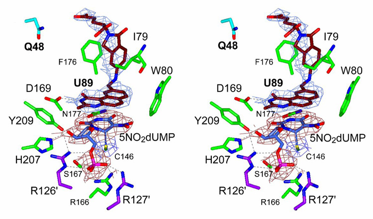
Fig. 8.
Stereo drawing of the active site of the crystal structure of the ternary complex of K48Q mutant TS with 5NO2dUMP and U89. Electron density is showed for nucleotide (red) and for antifolate (blue). Q48 is drawn with cyan bonds and other residues in the active site are colored in green. R126′ and R127′ from the other chain are colored in magenta. A covalent bond is present between C6 of nucleotide and C146 of TS. Hydrogen-bonds are represented with dashed lines.

