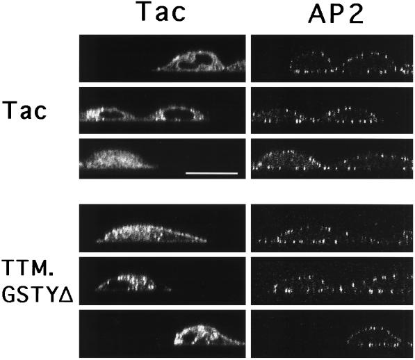
Figure 2.
Confocal microscopy ‘x-z’ images of Tac reporter expression and coated pit distribution in cells expressing chimeric receptors with tyrosine-based internalization signals. HeLa cells transiently transfected with Tac or TTM.GSTYΔ were stained as indicated in Figure 1, and vertical confocal ‘x-z’ images were collected of Tac (left panels) and AP2 (right panels). Note AP2 staining in both transfected and untransfected cells. Bar, 20 μm.

