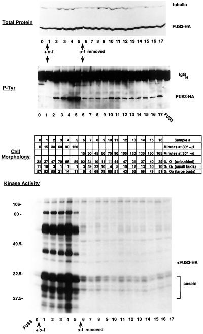
Figure 1.
Effect of α factor addition and withdrawal on Fus3-HA kinase activity. Strain EY960 (EY940 + pYEE121, FUS3-HA CEN URA3) was induced with α factor for 2 h, and then the α factor was washed out and the G1-arrested cells were allowed to recover for 3 h. Duplicate samples were taken at the indicated time intervals during the α factor addition and withdrawal; one sample was pelleted and frozen for cell extracts, and the other was fixed with formaldehyde for microscopic analysis. A total of 18 time points (0–17, 120 min in the presence of α factor, 180 min after α factor washout) were analyzed for protein levels, tyrosine phosphorylation, and kinase activity, and a total of 17 time points were analyzed for cell morphology (0–16, 120 min in the presence of α factor, 165 min after α factor washout). Fus3-HA was detected with 12CA5 antibody, and Fus3-HA tyrosine phosphorylation was detected with an anti-phosphotyrosine antibody as previously described (Elion et al., 1993). Kinase assays were performed as described (see MATERIALS AND METHODS). Top panel, Abundance of Fus3-HA by immunoblot analysis of 25 μg of whole-cell extract. Second panel, Tyrosine phosphorylation of Fus3-HA immunoprecipitated from 200 μg of whole-cell extract. Third panel, Cell morphology. Fourth panel, Fus3-HA kinase assay of associated substrates immunoprecipitated from 200 μg of whole-cell extract. Samples were separated on a 10% (38:2) acrylamide:bis-acrylamide SDS gel. Cell percentages are averages of three fields of 100 cells each.

