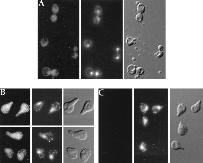
Figure 4.
Localization of Fus3-HA by indirect immunofluorescence. (A) FUS3-HA, vegetative growth; (B) FUS3-HA, 60-min α factor induction; (C) FUS3, 90-min α factor induction; (EY940 containing either FUS3 or FUS3-HA on a CEN plasmid [pYEE121]) grown either in the absence or presence of α factor, was prepared as described in MATERIALS AND METHODS and stained with 12CA5 antibody and affinity-purified donkey-anti-mouse IgG antibody conjugated to rhodamine-like Cy3. Micrographs shown are Cy3 fluorescence, DAPI fluorescence, and Nomarski differential interference contrast, as labeled. Cells were photographed with Fuji Super HGII-100 film using comparable exposure times.

