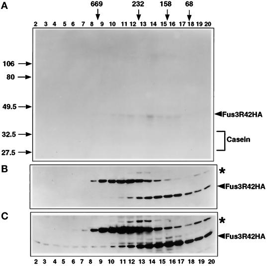
Figure 8.
Fus3R42 abundance and kinase activity across a 10–30% glycerol density gradient. (A) Fus3R42-HA kinase activity across the gradient assayed as described in Figure 6. (B) Distribution of Fus3R42-HA protein across the 10–30% glycerol density gradient, performed as in Figure 8. A portion of each fraction (50 μl) was used for immunoblot analysis to detect Fus3R42-HA with 12CA5 antibody. Strain EY940 containing FUS3R42-HA on a CEN plasmid was used for extract preparation. Cells were grown as in Figure 6. A similar exposure time is shown as in Figure 7B. (C) Longer exposure of the immunoblot in panel B showing Fus3R42-HA at the bottom of the gradient. The asterisk indicates a ∼55 kDa protein that cross-reacts with 12CA5.

