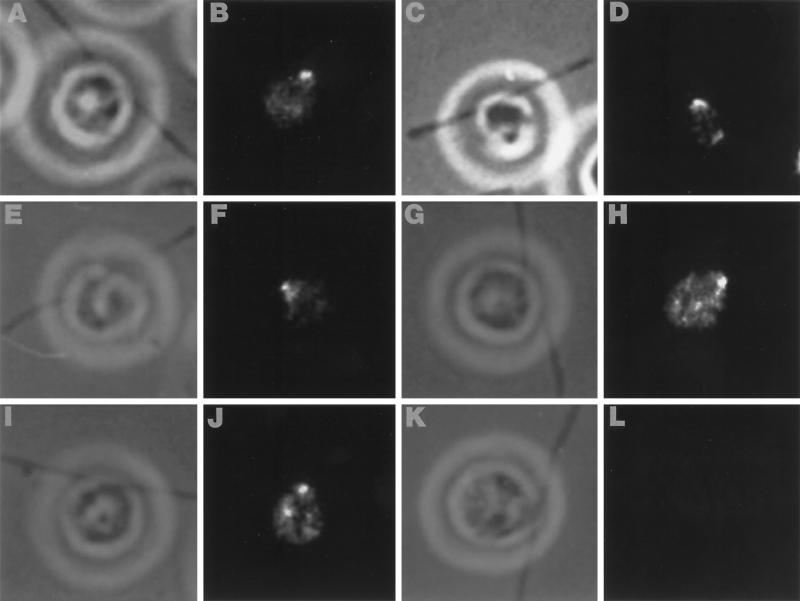
Figure 7.
Indirect immunofluorescence of whole cells demonstrates immunoreactivity near the basal bodies, but not along the length of the axoneme. Alternating phase contrast (A, C, E, G, and I) and anti-human p60 katanin fluorescence images (B, D, F, H, and J) of several different cells illustrating strong immunoreactivity at the base of the FBBC. Immune detection is with a FITC-conjugated secondary antibody. (K and L) Representative phase contrast and fluorecence images of a control cell that was treated with secondary antibody.

