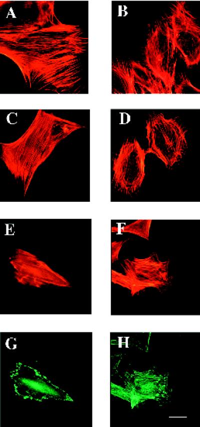
Figure 1.
SPARC-null mesangial cells exhibit an altered morphology in comparison to wild-type cells. Wild-type (A, C, E, and G) and SPARC-null mesangial cells (B, D, F, and H) were grown on coverslips in growth media. (A–F) BODIPY-conjugated phalloidin was used to visualize the actin cytoskeleton. (G and H) An anti-vinculin antibody was used to detect focal contacts in the mesangial cells. Bar, 5 μm.

