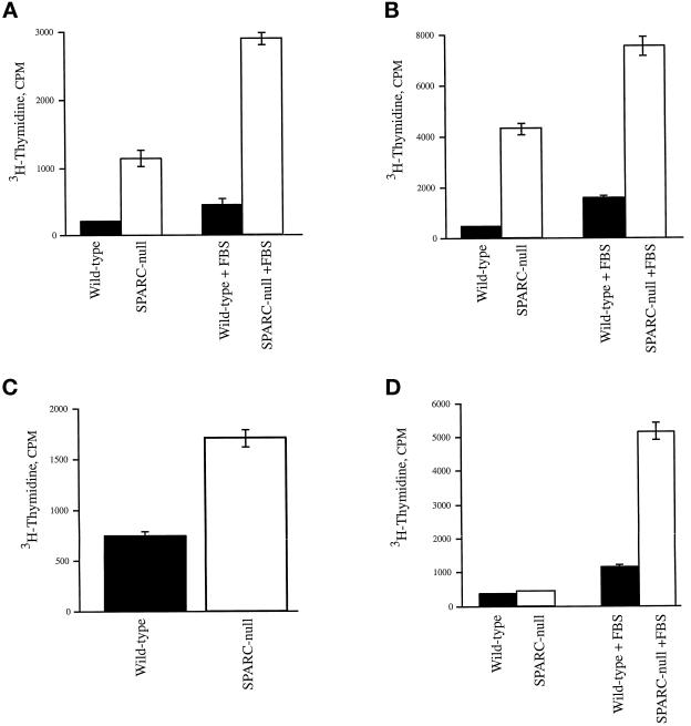
Figure 4.
SPARC-null primary cells incorporate higher levels of 3H-thymidine in comparison to wild-type cells. (A and B) Two separate preparations of wild-type (black bars) and SPARC-null mesangial cells (white bars) were assayed for incorporation of 3H-thymidine. Cells grown in resting media (see MATERIALS AND METHODS) for 24 h were treated with either 10% FBS in resting media or resting media alone for a further 18 h. 3H-Thymidine was added for the final 4 h of incubation. (C) Equal concentrations of wild-type (black bars) and SPARC-null fibroblasts (white bars) were plated in triplicate, in 24-well plates. The cells were grown in minimal media (1% FBS) for 24 h and subsequently stimulated with growth media (10% FBS) for 18 h. 3H-Thymidine was added for the final 4 h of incubation. (D) Wild-type (black bars) and SPARC-null aortic smooth muscle cells (white bars) were plated at equal concentrations in serum-free media for 48 h and either stimulated with 10% FBS or left in serum-free media for an additional 18 h, as described above for mesangial cells. 3H-Thymidine was added for the final 4 h of incubation. Error bars represent the SEM.

