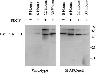
Figure 6.
Basal levels of cyclin A are increased in SPARC-null versus wild-type mesangial cells. Both cell types were plated at equal concentrations and grown in resting media for 48 h before the addition of 10 ng/ml PDGF. Cells were harvested in RIPA buffer with protease inhibitors at 6, 12, and 30 h. Equal amounts of protein were loaded in each lane, separated on a 10% SDS-PAGE gel under reducing conditions, transferred to nitrocellulose, and probed with an anti-cyclin A antibody. Immunoreactive bands were detected by enhanced chemiluminescence. Equal loading of protein was confirmed by staining of the transferred gel with Coomassie blue. Molecular weight markers (kilodaltons × 10−3) are indicated. The identity of the band at Mr 75,000 is unknown.

