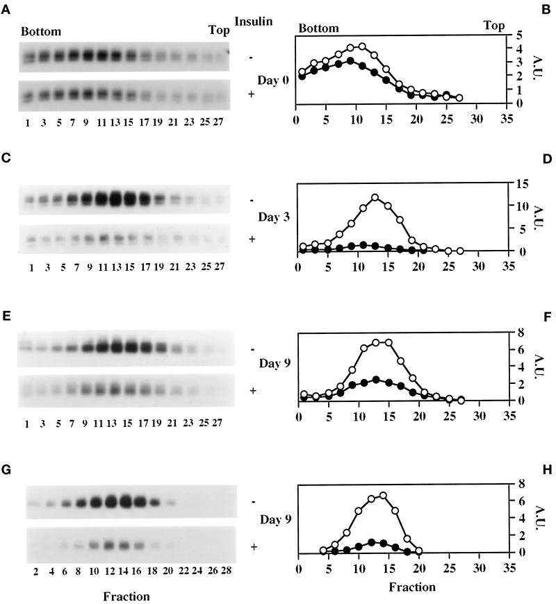
Figure 4.
By day 3, GLUT1 resides in a distinct and fully insulin-responsive compartment, which cosediments with GLUT4-containing vesicles from differentiated cells. On days 0 (A and B), 3 (C and D), and 9 (E-H), 3T3-L1 cells were serum starved for 2 h before treatment with or without 100 nM insulin for 30 min. Light microsomes (1.5–2 mg) were then isolated and sedimented in a 4.6-ml 10–30% sucrose gradient as described in MATERIALS AND METHODS. The panels on the left show the Western blots of odd- and even-numbered gradient fractions, which were immunoblotted for GLUT1 and GLUT4, respectively. Detection was with HRP-conjugated secondary antibodies and chemiluminescence. These data were quantified by densitometry and are graphically displayed (circles) in the panels on the right as arbitrary units (A.U.). Open and closed symbols, basal and insulin-stimulated conditions, respectively. A representative profile of the total protein in these gradients is shown in Figure 6E. These data are representative of three independent experiments.

