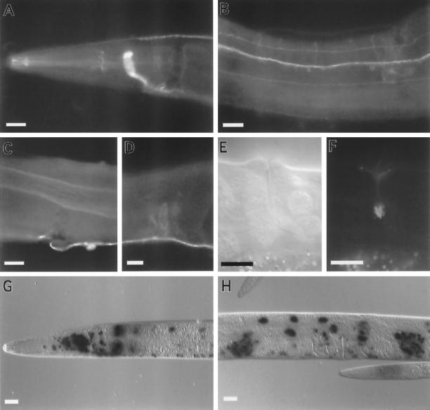
Figure 2.
Expression of unc-64 syntaxin. (A–D) Whole adult wild-type hermaphrodite worms fixed and stained with α-UNC-64 primary antibodies and visualized with FITC-conjugated antibodies. (A) Lateral view of the head region showing immunoreactivity in the nerve ring, dorsal cord, and pharyngeal nervous system. (B) Ventral view of the midbody region showing expression in ventral cord axons, neuronal cell bodies, and in commissural and sublateral processes. (C) A lateral view of the midbody showing UNC-64 immunoreactivity on the basolateral surface of the intestine. (D) A lateral view of the midbody showing immunoreactivity in the hermaphrodite spermatheca. The male vas deferens also stains. (E) A lateral view of the vulva viewed using differential interference contrast microscopy. (F) Same plane of focus as panel E showing fluorescence from the UNC-64 A::GFP product in the uv1 cells of the vulva. (G–H) Whole adult wild-type animals fixed and stained for LacZ gene activity expressed under the control of the unc-64 promoter. LacZ activity assayed using X-gal as a substrate. The lacZ gene contains a nuclear localization signal. (G) Lateral view of the head region showing expression of unc-64 ::lacZ in neuronal and intestinal nuclei. (H) Lateral view of the midbody of an adult hermaphrodite showing unc-64::lacZ expression in intestinal nuclei, neuronal nuclei, and the spermatheca. Scale bar, in all panels, 20 μm.

