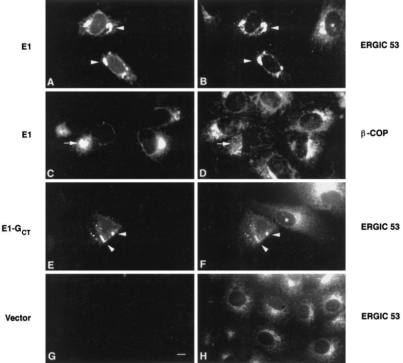
Figure 6.
ERGIC 53, but not β-COP, colocalizes with E1 and E1-GCT in transiently transected Vero cells. Vero cells were transiently transfected with expression plasmids encoding rubella E1 or E1-GCT and processed for indirect immunofluorescence after 40 h as described in Figure 3. E1 and E1-GCT were visualized using human anti-E1 serum and FITC-goat anti-human IgG (panels A, C, E, and G). Monoclonal antibodies to ERGIC 53 and β-COP and Texas Red-goat anti-mouse IgG were used in panels B, D, F, and H as indicated. In cells expressing E1 and E1-GCT, the majority of ERGIC 53 was present in the tubular pre-Golgi compartment (A, B, E, and F, arrowheads), whereas in untransfected cells it was concentrated in a perinuclear vesicular pattern in the Golgi region (B, F, and H, asterisks). In contrast, staining for E1 and β-COP (C and D, arrows) or E1-GCT and β-COP (our unpublished observations) did not overlap significantly. In panel G, cells were transfected with vector alone to show that the human anti-E1 serum did not cross-react with endogenous ERGIC 53. Bar, 10 μm.

