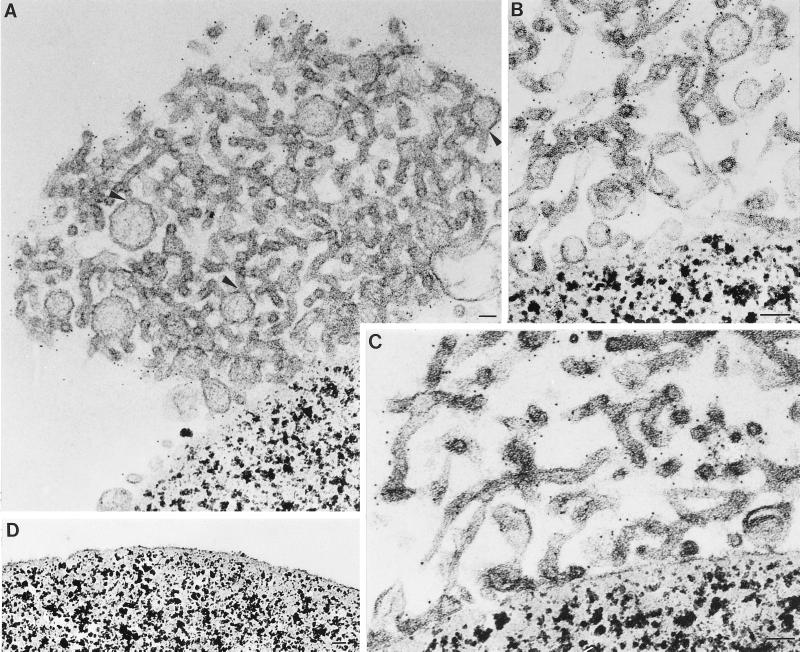
Figure 8.
Isolated tubular networks contain vesicles and budding structures. (A–C) Immunoisolated tubular networks isolated from BHK-E1GCT cells were processed for EM while still attached to P5D4-coated magnetic beads. Vesicles and budding structures that are associated with the tubular nests are evident in panels A and B. In panel A, regions of apparent continuity between budding structures and the tubules are indicated by arrowheads. Samples were incubated with a rabbit antibody directed against the VSV G epitope on E1-GCT followed by 5 nm gold coupled to protein A. Labeling is confined to the cytoplasmic surface of the tubules. (D) Beads coated with an irrelevant monoclonal antibody (BW8G65) were incubated with smooth membrane fractions prepared from BHK-E1-GCT cells and prepared for EM as described above. No smooth membranes are seen attached to the beads. Bar, 50 nm in all panels.

