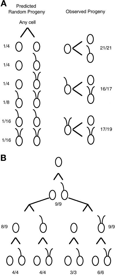
Figure 2.
Pedigree analysis of the mitotic progeny from uni3–1 cells. Individual cells were placed in 40 μl rich medium in microtiter wells. Upon mitotic cell division, each daughter cell was transferred into a new well, and the swimming phenotype was monitored under a dissecting microscope with 100× magnification. (A) In the left panel, the predicted fraction of pairs of daughter cells if the phenotype is random and independent of lineage. The fractions were calculated as simple probabilities for independent events. In the right panel are the observed numbers. The fractions indicate the number of cells with the diagrammed phenotype over the number of cells successfully transferred and observed. In the three pairs that did not follow the pattern diagrammed, both cells had no flagella when observed. (B) Twelve aflagellate cells were placed individually in microtiter wells, and progeny for three successive cell divisions were monitored. The fractions indicate the number of cells that showed the diagrammed pattern over the number successfully transferred and monitored. As in part A, cells that did not show the diagrammed pattern produced two aflagellate cells.

