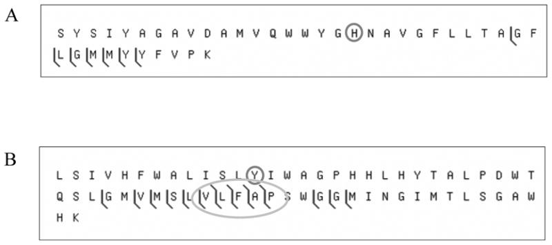
Figure 3.

CAD fragmentation of the crosslinked tryptic peptide from the C-type oxygen reductase assuming the absence of the crosslink. The CAD fragmentation dataset used to generate figure 2 was also analyzed as if the crosslink were not present. (A) shows the matches for the tryptic peptide S193-K232 and (B) for L243-K304. The histidine and tyrosine normally involved in the crosslink are circled. The oval highlights several fragments that were found to span the crosslinked residue but do not include the mass of the opposite crosslinked peptide.
