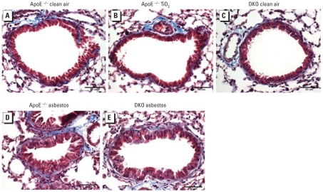
Figure 2.
Representative lung sections stained with Masson’s trichrome after 30-day exposures, showing inflammatory cell infiltration and fibrosis in asbestos-exposed animals. Blue indicates collagen associated with fibrosis. (A) ApoE−/− mice exposed to clean air. (B) ApoE−/− mice exposed to TiO2. (C) DKO mice exposed to clean air. (D) ApoE−/− mice exposed to asbestos. (E) DKO mice exposed to asbestos.

