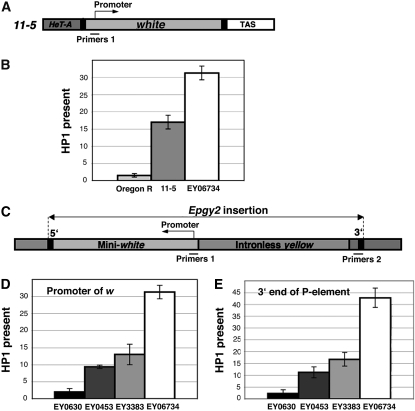
Figure 3.—
HP1 is found at telomeric insertions. (A) 11-5 bears a complete white gene inserted between the terminal retrotransposon array and TAS at the 2L telomere. Primers 1 used in the ChIP experiment surround the promoter. (B) ChIP analysis of HP1 at the w promoter of Oregon R, 11-5, and EY06734. Graphs represent real-time PCR results obtained after ChIP. HP1 measurements were normalized to 5% of input DNA and further normalized to the RpL32 locus. Error bars represent standard deviations. (C) The EPgy2 construct of EY insertions has a mini-white gene (mini-w) and an intronless yellow gene. Primers 1 and Primers 2, which correspond to the mini-w promoter and the 3′ end of the P-element insertion, respectively, were used for PCR analysis after ChIP. (D and E) The level of HP1 binding at the w promoter (D) and the 3′ end of the P element of EY insertions (E) analyzed by ChIP. Error bars represent standard deviations.

