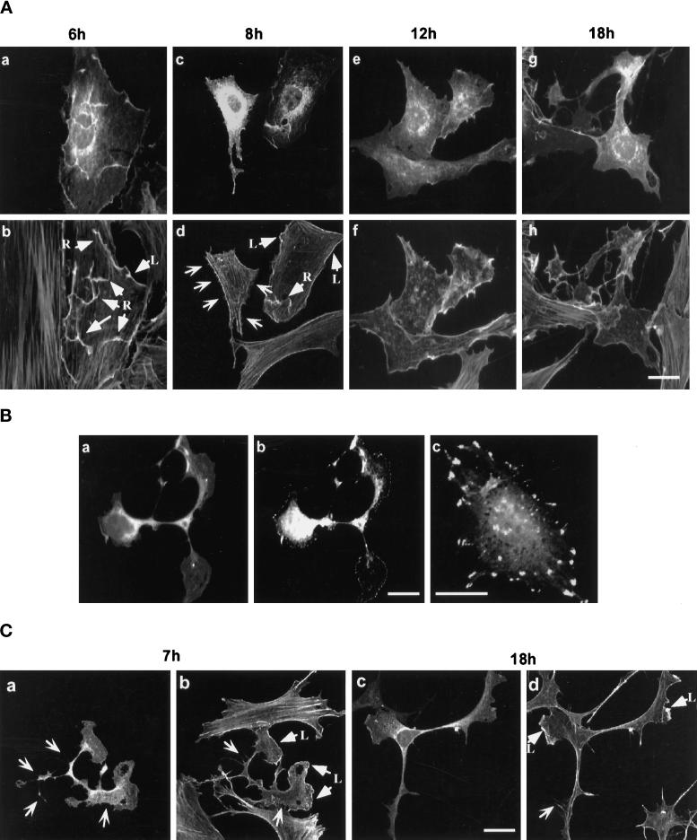
Figure 2.
Actin reorganization induced by GFP-RhoGV12 protein expression in REF-52 and Swiss 3T3 cells. (A) Quiescent REF-52 cells were microinjected with plasmids encoding GFP-RhoGV12 protein. Microinjected cells were fixed after different periods of time and then stained with rhodamine-labeled phalloidin. Cells were observed under confocal laser scanning microscopy for GFP activity (panels a, c, e, and g) and for filamentous actin distribution (panels b, d, f, and h). Arrows designated with an R and L indicate dorsal ruffles and lamellipodia, respectively, whereas open arrows indicate filopodia. Bar, 10 μm. (B) Exponentially growing Swiss 3T3 cells were transfected with constructs expressing GFP-RhoGV12 protein and serum starved for 14 h. Cells were fixed and monitored for GFP activity (panel a) and phosphotyrosine-containing epitopes (panels b and c). (C) Exponentially growing Swiss 3T3 cells were transfected with constructs expressing GFP-RhoGV12 protein. Cells were either fixed 7 h (panels a and b) or 18 h (panels c and d) after transfection and stained with rhodamine-labeled phalloidin. Cells were monitored for GFP activity (panels a and c) and filamentous actin (panels b and d). Arrows designated with an L indicate lamellipodia whereas open arrows indicate filopodia. For each panel, cells shown are representative of more than 100 observed cells.

