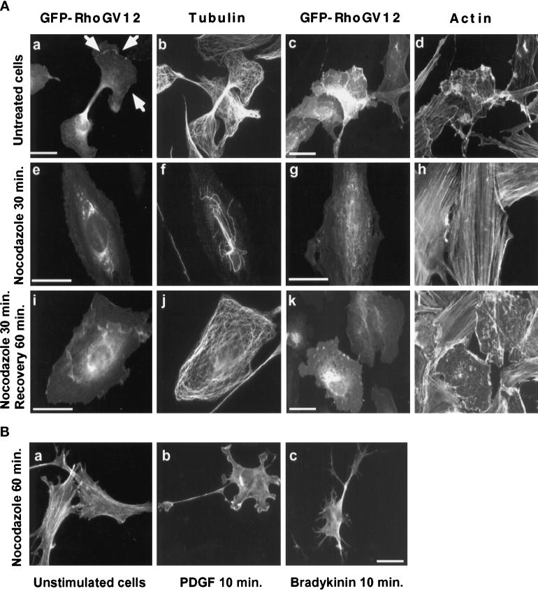
Figure 9.
Effect of microtubule depolymerization on GFP-RhoG protein localization and activity. (A) REF-52 cells were transfected with constructs expressing GFP-RhoGV12 protein. Eighteen hours later, transfected cells (panels a–d) were treated for 30 min with 2 μM nocodazole (panels e–f). Cells were then rinsed and incubated for 60 min in media without nocodazole (panels i–l). Cells were fixed and monitored for GFP activity (panels a, c, e, g, i, and k), microtubule distribution (panels b, f, and j), and F-actin distribution (panels d, h, and l). Bar, 10 μm. (B) Swiss 3T3 cells were treated for 60 min with 2 μM nocodazole (panel a) and then stimulated for 10 min with 3 ng · ml−1 PDGF (panel b) or 100 ng · ml−1 bradykinin (panel c). Cells were fixed and observed for F-actin distribution. For each panel, cells shown are representative of more than 100 observed cells.

