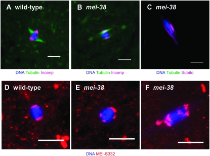
Figure 2.—
Localization of spindle- and centromere-associated proteins in stage 14 oocytes. Wild-type oocytes (A and D) and mei-381 mutant oocytes (B, C, E, and F) are shown. Oocytes were stained for Incenp (A and B), Subito (C) (all in magenta), and MEI-S332 (red). Subito and Incenp show identical staining patterns in wild-type oocytes (Jang et al. 2005), thus only a wild type with Incenp staining (A) is shown here. Tubulin staining is green in A and B and DNA is in blue. Note the uneven distribution of MEI-S332 signals in B even though the karyosome looks normal. Bar, 5 μm.

