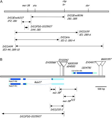
Figure 3.—
Genetic mapping and cloning of mei-38. (A) Deficiencies (arrows) in the mei-38 region shown relative to the genetic map (top line). The cytological breakpoints of each deficiency are indicated at the bottom of its name. (B) Physical map of the mei-38 region. Triangles mark the positions of each transposon insertion. The blue boxes show the transcription units with the coding regions in dark blue and start and stop codons shown with green and red vertical lines, respectively (from Gelbart et al. 1997). At the bottom are the two regions deleted in the original mei-381 mutant and three deletions generated in this study. Df(1)FDD-0225927 was generated through recombination between two FRT sites, one in P{XP}d04500 and the other in PBac{RB}e04351, located ∼10 kb distal to Rab27.

