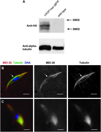
Figure 4.—
Localization of MEI-38. (A) Western blot of ovary protein from females expressing HA-tagged MEI-38 protein or wild-type females using antibodies to the HA epitope or α-tubulin as a control. (B) Immunolocalization of MEI-38 (red) relative to tubulin staining (green) and DNA (blue) in stage 14 oocytes. MEI-38 colocalizes with microtubule staining except in the central spindle (arrow). (C) MEI-38 colocalizes with all microtubule staining in sub1/sub131 oocytes. Bar, 5 μm.

