
Figure 3.
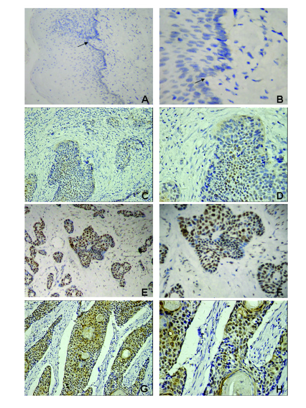
Expression analysis of CENP-H protein by immunohistochemistry. CENP-H expression was mainly localized within nuclei of tumor cells, and diffuse staining was observed in some tumor cells. CENP-H is not expressed in normal epithelial cells. A and B Staining of CENP-H in normal esophageal epithelial tissue (arrow, normal epithelial cells). C and D, low expression of CENP-H in esophageal carcinoma tissues (200 and 400, respectively). E, F, G and H, high expression of CENP-H in esophageal carcinoma tissues (200 and 400, respectively).