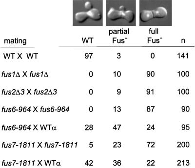
Figure 1.
Defects of the cell fusion mutants. Filter matings were analyzed microscopically using DIC optics in combination with DAPI, a fluorescent DNA marker. The classes of zygotes scored were WT, which had no septum at the zone of fusion and a fused nucleus; partially defective (partial Fus−), which showed an obvious cell fusion septum and a fused nucleus; and completely defective (full Fus−), which had a pronounced septum and unfused nuclei. The percentages of each type of zygote are shown. The far right column shows the number of zygotes scored. The counting error is within 5%. The matings used in this analysis were WT × WT (MY3371 × MY2787), fus1Δ × fus1Δ (MY4161 × MY4164), fus2Δ × fus2Δ (JY424 × JY428), fus6-964 × fus6-964 (MS2680 × MS2679), fus6-964 × WT (MS2680 × MS2104), fus7-1811 × fus7-1811 (MY4477 × MY3785), and fus7-1811 × WT (MY4477 × MY2787). The photographs of the DIC–DAPI double images and the data from the mating between MY4477 × MY3785 have been reported elsewhere (Brizzio et al., 1998).

