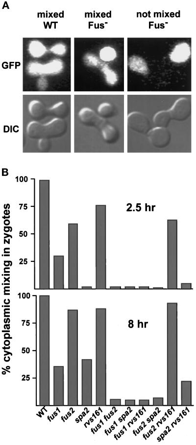
Figure 2.
Cytoplasmic mixing in zygotes using a soluble fluorescent marker, GFP. (A) Three representative types of zygotes seen in the cytoplasmic mixing assay. The zygotes with mixed cytoplasms consisted of those with no obvious zone of cell fusion remaining (mixed WT) and zygotes with a cell fusion defect (mixed Fus−). Prezygotes with no removal of the intervening cell wall (not mixed Fus−) were also scored. The percentages of mixed zygotes (includes the mixed WT and the mixed Fus−) are shown in B. For each mating, a minimum 100 zygotes were counted. The counting error is within 5%. The matings were as follows: WT (MY3371 × MY2787), fus1 (JY427 × MY4164), fus2 (JY424 × JY428), spa2 (MY3608 × MY3773), rvs161 (MY3909 × MY4495), fus1 fus2 (MY4160 × JY429), fus1 spa2 (MY4817 × MY4819), fus1 rvs161 (MY4905 × MY4907), fus2 spa2 (MY5067 × MY4815), fus2 rvs161 (MY4801 × MY4802), and spa2 rvs161 (MY5062 × MY4748).

