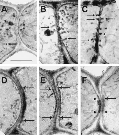
Figure 6.
Electron-dense plaques and invaginations at the zones of cell fusion in fus2 and rvs161 prezygotes. The micrographs are from matings of rvs161-P203Q (MY5224 × MY5359) (A), rvs161▵ (MY3909 × MY4495) (B, C, and F), and fus2 (MY4158 × MY4178) (D and E). Examples of invaginations (A–C) are accentuated with arrows. The electron-dense plaques are along the plasma membrane between the arrows in D–F. Bar, 0.5 μm.

