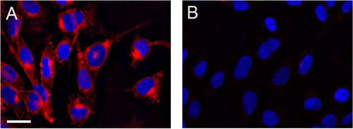
Figure 4.

Fluorescence microscopy of CHO cells labeled with 1. Cells incubated for 3 d in the presence (A) or absence (B) of Ac4ManNAz (100 μM) were treated with 1 (200 μM) for 2 h at 37 °C. The cells were then fixed and permeabilized with MeOH and stained with DAPI before imaging. Red = Cy5.5 channel. Blue = DAPI channel. Scale bar = 20 μm.
