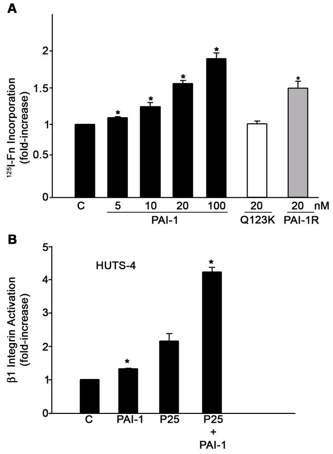
Fig. 1.
PAI-I increases fibronectin matrix assembly and β1 integrin activation in MG-63 cells. A - MG-63 cells were incubated with 125I-fibronectin for 6 hours in the presence of the indicated concentrations of PAI-1, PAI-1R or PAI-1 (Q123K) in DMEM+BSA 0.02% + 20 mM Hepes. Cell layers were extracted with 1% DOC, and 125I-fibronectin that was incorporated into the detergent-insoluble matrix was recovered by centrifugation and measured by gamma scintillation. This experiment is representative of three separate experiments. Data are the mean ± S.E. of two experiments performed in duplicate. *Significantly different than control cells, t test, p < 0.05 (n=4). B - MG-63 cells were pretreated with 20 nM PAI-1 for 20 minutes and incubated with 50 μM of uPAR agonist P25 or S25 for 1 hour in DMEM. Activation of β1 integrin was assessed by ELISA using the HUTS-4 antibody. Total β1 integrin was measured using the P5D2 antibody against α5β1. The graph shows the levels of β1 integrin activation after normalization to total β1 levels. Neither P25 nor PAI-1 had any effect on total β1 integrin. Data represent one of three different experiments performed in triplicate. *Significantly different than control or P25-treated cells, respectively, t test, p < 0.05 (n=3).

